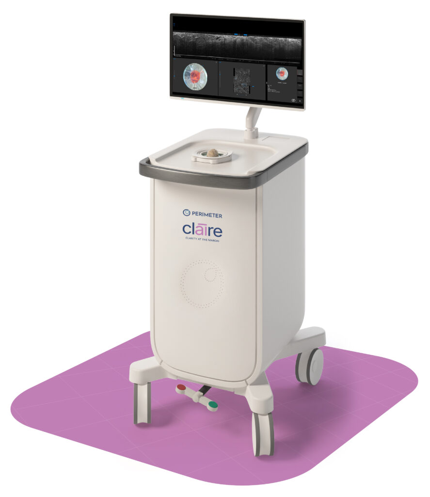
Bringing clarity to margin status when it matters most.Bringingclarity tomarginstatus
whenit mattersmost.
See the full picture with PerimeterSeethefullpicture with Perimeter.
See the full picture with Perimeter.
Groundbreaking Margin Visualization
Perimeter OCT Technology
Harnessing the power of advanced technology and intuitive functionality, Perimeter Medical Imaging is driven to transform cancer surgery with advanced imaging tools. The Perimeter Optical Coherence Tomography (OCT) system provides cross-sectional, real-time margin visualization of tissue microstructures during surgical procedures. Perimeter’s game-changing technology gives surgeons unmatched clarity and confidence on margins at the point of care.
Using light, images have 10x higher resolution than ultrasound and X‑ray and 100x higher than MRI for visualization at the cellular level.
Resolution is optimized for visualizing microscopic tissue structures down to 2mm, ideal for reviewing surgical margins.
Tissue is entirely preserved for histopathology and device does not come in contact with the patient or enter the sterile field.
Identify and annotate regions of interest with image review manipulation tools for optimized decision-making in the operating room.
Label and capture images of surgical margins on all six planes to convey accurate orientation for pathology.
With the Perimeter S-Series OCT system, breast surgeons have a much better chance of getting clean margins the first time we do a lumpectomy.
Michele Carpenter, MD, FACS
I believe this technology represents the next standard of care in cancer surgery.
Amelia Tower, DO
In the OR, Perimeter’s technology provides ultra-high resolution visualization of my specimen in real-time. This adds to my confidence on the final margin status.
Beth DuPree, MD, FACS, ABOIM
Watch our symposium at the American Society of Breast Surgeons Annual Meeting
In this educational session, you’ll learn how Perimeter’s margin visualization technology gives surgeons the real-time insights they need to personalize clinical decisions at the point of care.
Featured
News
Perimeter announces first commercial placement of its S-Series OCT in the state of Georgia.
Webinar Replay
Getting clean margins remains a challenge for surgeons. Hear from breast surgeons Dr. Michele Carpenter, who described OCT as “like having a pathology slide in the OR,” and Dr. Beth DuPree, an early OCT user.
Case Study
Breast cancer surgeon, Dr. Beth DuPree, explains the benefits of using Perimeter's S-Series OCT technology in the OR.
Case Study
Dr. Amelia Tower, breast surgical oncologist, describes how she used Perimeter’s OCT imaging in the OR to evaluate tissue microstructures and identify regions of concern that were not detected with specimen radiography.
Patient Story
Nadia shares her journey with breast cancer and infertility, as well as her treatment plan with Perimeter's technology.
Clinical Paper
This study's findings suggest that WF-OCT may be a promising imaging modality for intraoperative analysis in head and neck surgery, especially at deep margins, without impacting specimen integrity or surgical and pathology workflows.