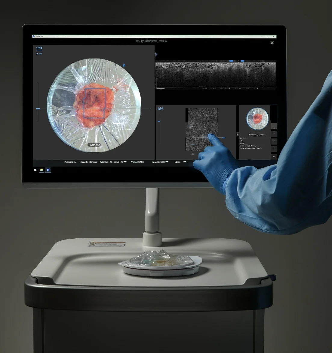
Let’s walk through our latest technology, Claire, an AI-enabled OCT device. We will learn how Claire
works, how it is used by your surgeon to assess tumor margins during breast conserving surgery,
and how this could prevent the need for a second surgery.

We’ve also included some downloadable resources that you can take to your appointment to be more prepared.
As you navigate your breast cancer, we hope you find these helpful and encouraging.

EXPLORE MORE

WHAT ARE MARGINS

What are surgical margins and why do they matter during a lumpectomy?

Your surgeon will remove the cancerous tumor and some normal tissue—the margin—that surrounds it. After the procedure, a pathologist will carefully examine the tissue to see if cancer cells are present in the margins which could indicate that cancer may have been left behind in the surgical site. This report typically takes 2-7 days to get back.

icon

Guidance on Margins

Different types of breast cancer require different approaches. Your surgical team follows established medical guidelines to make sure they remove the right amount of tissue for your specific diagnosis.

Margins that are free of cancer are called "negative" or "clear" margins. If the excised tissue contains cancer cells at the outer edge, it is considered "positive." Some types of cancers require a small rim--about 2mm, or the thickness of a nickel--of healthy tissue surrounding the tumor to also be excised.

icon

The Challenge of DCIS

Certain types of breast cancer, such as Ductal Carcinoma in Situ (DCIS), can be especially challenging to see.

Because DCIS is microscopic and often cannot be felt by the surgeon, it can be especially challenging to detect while operating because it often feels and looks the same as healthy tissue.

icon

Here’s why
margins matter

Clean (or clear) margins are linked to better outcomes and lower cancer recurrence. 1

If the pathologist determines that the tumor margins are positive or close, your surgeon may recommend second surgery, called a re-excision, to reduce the likelihood of recurrence.

This could be another lumpectomy (re-excision), or, in some cases, a mastectomy.

icon

Microscopic
Clarity in the OR

During your lumpectomy, your surgeon uses a variety of imaging tools and will visually inspect and feel the tissue to determine where the cancer is located.

Imaging technologies, such as Perimeter’s Claire, may be used during surgery to support your surgeon’s evaluation of the removed tissue allowing your team to see 10x more detail than traditional imaging while you are still in the operating room.

After your surgery, the lumpectomy specimen will be sent to pathology for final analysis and evaluation.

1. Ezzat A, Shanthakumar D, Laskar N, et al. Impact of resection margin width on local recurrence following breast-conserving surgery and whole breast
radiotherapy for pure ductal carcinoma in situ: a systematic review and meta-analysis. BMJ Oncol. 2025;4(1):e000633. Published 2025 Mar 6.
doi:10.1136/bmjonc-2024-000633

During your surgery, Claire presents
your surgeon with microscopic images
of removed tissue. This additional insight
has the potential to prevent subsequent
surgeries due to cancer left behind.

icon

Real-time Clarity for Your Choice

If you choose a lumpectomy, advanced imaging tools like Perimeter’s Claire provide real-time clarity during your procedure.

By providing an AI-enabled ‘extra set of eyes’ to evaluate the removed tissue while you are still in the operating room, this technology offers additional information to support your team’s goal of preserving the breast.

icon

Resource: Example Questions to Ask Your Surgeon

We created a downloadable list of example questions that you can take with you to your surgery appointment.

We hope that this list can help you feel more prepared as you go through your decision-making process.

SEE THE LIST

AI-Enabled, real-time, 3D imaging of the full 2mm margin.

Claire™ OCT brings additional precision to your surgery. Here is how this advanced tool works to give your surgical team new insights:

How OCT Works

Think of Optical Coherence Tomography (OCT) as an “ultrasound with light.” It works similarly to a traditional ultrasound, but it uses light instead of sound to create a high-definition 3D scan of microscopic details with much higher resolution.

10x Clearer than X-ray

This advanced imaging allows your surgical team to see details on a lumpectomy specimen 10 times clearer than a standard X-ray.

Real-Time Insight

Because this imaging happens right in the operating room, it is designed to help your surgeon see intricate details along the margins while you are still in surgery.

An AI-enabled “Extra Set of Eyes”

Claire also uses AI software to help flag specific areas for the surgeon to prioritize for review. This added layer of information gives your team the potential to make informed, real-time decisions before your surgery is even finished.

Claire combines advanced Optical Coherence Tomography (OCT) with an AI-enabled image analysis tool to bring real-time 3D clarity
to the microscopic level across the full 2mm margin. Claire has been clinically validated to highlight regions within the OCT image that
are suspicious for breast cancer in order to quickly guide attention to the most concerning regions.2

AI overlays can be toggled
ON, Show Less, or OFF

Highlights suspicious microstructural
features associated with DCIS & IDC

Prioritized review with
look-here assistance

Supports
surgeon-led review

SAY HELLO TO CLAIRE AI+OCT

2. Perimeter Medical Imaging AI. Wide Field OCT + AI for Positive Margin Rates in Breast Conservation Surgery. Identification No NCT05113927. Last
updated December 12, 2024. Available at: https://clinicaltrials.gov/study/NCT05113927.

2. Perimeter Medical Imaging AI. Wide Field OCT + AI for Positive Margin Rates in Breast Conservation Surgery. Identification No NCT05113927. Last updated December 12, 2024. Available at: https://clinicaltrials.gov/study/NCT05113927.

Claire Intended Use/Indications for Use

The Claire OCT System’s indications for use are as an adjunctive three-dimensional imaging tool which provides volumetric cross-sectional, real-time depth visualization, coupled with an artificial intelligence computer aided detection algorithm which identifies and marks focal areas suspicious for breast cancer and is used concurrently with physician interpretation of the images. The Claire OCT System is intended for use in conjunction with other standard methods for evaluation of the margins of excised lumpectomy tissue during surgical procedures in patients with a biopsy-confirmed diagnosis of breast cancer.

Limitations of Use

  • concurrent use in surgeries with cryo-assisted localization
  • use with specimen margins that have been destroyed, damaged, or are otherwise not intact prior to imaging.

This device has not been evaluated for individuals who:

  • Are under the age of 18
  • Are male
  • Have metastatic cancer (Stage IV)
  • Have lobular carcinoma as their primary diagnosis
  • Have had previous ipsilateral breast surgery for benign or malignant disease within 2 years (including implants and breast augmentation)
  • Have multi-centric disease (histologically diagnosed cancer in 2 different quadrants of the breast), unless resected in a single specimen
  • Have bilateral disease (diagnosed cancer in both breasts)
  • Are currently lactating
  • Are currently pregnant

Contraindications of Use

  • The Claire OCT System should not be used to replace standard tissue histopathology assessment.
  • The Claire OCT System should not be used for diagnosis.
rx-icon

Federal law restricts this device to sale by or on the order of a physician.

rx-icon

The Claire OCT System device offers an additional source of information to the user by providing rapid, automated visualization of near surface microstructure. This information is an adjunct to visual observation of the surface of an excised tissue sample and other intraoperative margin assessment techniques.