Perimeter Medical Imaging AI intends the sharing of this content for an investor audience and not for use by healthcare professionals. The data contained herein have not undergone peer review nor evaluation by FDA and should not be used to guide clinical practice. The S-Series OCT has 510(k) clearance under a general indication and has not been evaluated by FDA specifically for use in breast tissue, breast cancer, other types of cancer, margin evaluation, and reducing re-excision rates. For more information, visit https://perimetermed.com/disclosures or contact medicalaffairs@perimetermed.com.
Perimeter Claire™ OCT is an FDA-approved, adjunctive AI-enabled imaging tool. It is intended for use in evaluating margins of excised lumpectomy tissue and does not replace standard histopathology. Perimeter’s Claire OCT is not available for sale outside the USA.
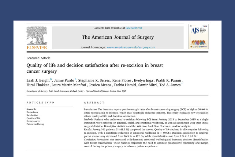
This study evaluates how re-excision following breast conserving surgery (BCS) affects quality-of-life and decision satisfaction. It highlights that re-excision is associated with a signifi cant decline in emotional wellbeing and patient satisfaction with lumpectomy. It also recommends that strategies to improve margin control during the initial surgery help reduce the risk of re-excision and preoperative counseling may help align patient expectations and enhance experience with surgery.
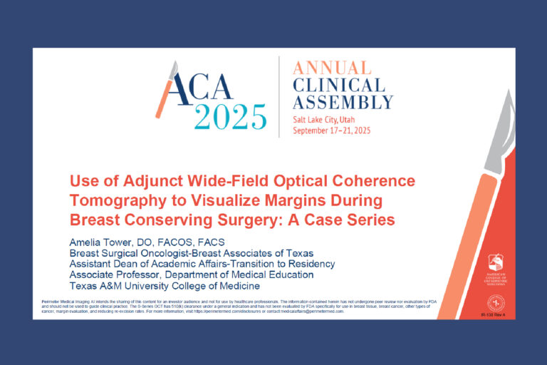
Dr. Amelia Tower recently presented her case series at the 2025 Annual Clinical Assembly of Osteopathic Surgeons by American College of Osteopathic Surgeons. Her presentation discusses case series where the breast surgical oncologist, used OCT for tissue visualization at the point of care, saving three patients with DCIS from a repeat surgery. She highlighted Perimeter’s advanced imaging technology and its impact on surgical precision and patient care.
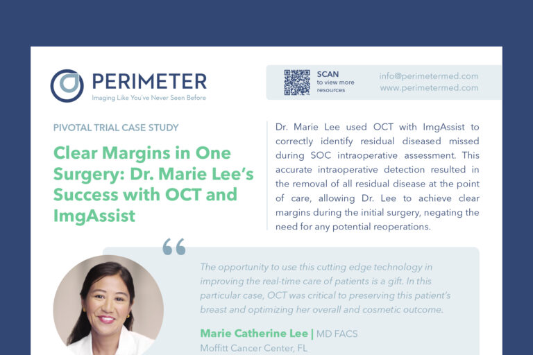
Dr. Marie Lee used OCT with ImgAssist to correctly identify residual diseased missed during SOC intraoperative assessment. This accurate intraoperative detection resulted in the removal of all residual disease at the point of care, allowing Dr. Lee to achieve clear margins during the initial surgery, negating the need for any potential reoperations.

This paper provides an analysis on how age and disease type influence reoperation rates after breast-conserving surgery, revealing a downward trend with increasing age and significant differences across disease types.

This paper concludes that the use of intraoperative OCT imaging in breast-conserving surgery can significantly reduce reoperation rates by improving margin assessment, suggesting that OCT may serve as a valuable tool to help achieve negative margins during primary surgery.

This paper comprehensively analyzes current reoperation rates for breast-conserving surgery patients and their associated implications, underscoring the need for new strategies and technologies to address this problem.

Amelia Tower, DO, FACOS; Mar. 21, 2023
Three women with biopsy confirmed DCIS and/or IDC undergoing breast-conserving surgery had their lumpectomy tissue imaged using intraoperative specimen radiography followed by WF-OCT. The surgeon used WF-OCT imaging in the OR to evaluate tissue microstructures and identify regions of interest that were not detected with intraoperative specimen radiography, allowing the surgeon to make real-time clinical decisions to excise additional tissue during the primary surgery. Pathology confirmed that all final margins were negative for residual disease. The removal of the additional tissue saved these 3 patients from the need for a second surgery.

Youngran Kim, PhD; Cecilia Ganduglila-Cazaban, MD, DrPH; Nina Tamirisa, MD; Anthony Lucci, MD; Trudy Millard Krause, DrPH; Annals of Surgical Oncology, Feb. 06, 2024
This study was designed to provide a comprehensive and up-to-date understanding of population-level reoperation rates and healthcare costs associated with reoperation for patients who underwent breast-conserving surgery (BCS). The rates of reoperation after BCS have remained high and have contributed to increased healthcare costs.

Yanir Levy, David Rempel, Mark Nguyen, Ali Yassine, Maggie Sanati-Burns, Payal Salgia, Bryant Lim, Sarah L. Butler, Andrew Berkeley, Ersin Bayram; Life-Optical Imaging and Fluorescence Imaging in Breast Cancer Diagnosis and Surgery. Dec. 14, 2023
This study explores the integration of Wide Field Optical Coherence Tomography (WF-OCT) with an AI-driven clinical decision support system, with the goal of enhancing productivity and decision making in breast cancer margin assessment. The results suggest the investigational deep learning model accurately identified 96.8% of pathology positive margins in WF-OCT images with high sensitivity and specificity.

Accurate assessment of tumor size is crucial for effective surgical planning in cancer patients. Specific to breast cancer, however, preoperative imaging with mammography and ultrasound often underestimates the actual tumor size. This paper examines the drivers of underestimation, discusses clinical implications, and reviews methods to minimize the negative impact of underestimation on surgical outcomes.

Beryl Rabindran, Adriana D. Corben; Pathology and Oncology Research, Jul. 14, 2023
Various tissues samples were obtained from a single autopsy and were imaged with WF-OCT then processed for permanent histology. The WF-OCT images captured in this study displayed the key features of the most common human tissue types encountered in surgical oncology with utility comparable to histology, confirming the utility of an FDA-cleared imaging platform. With further study, WF-OCT may have the potential to bridge the gap between the immediate information needs of the operating room and the longer timeline inherent to histology workflow.

Arvind K. Badhey, Julia S. Schwarz, Benjamin M. Laitman, Brandon M. Veremis, William H. Westra, Mike Yao, Marita S. Teng, Eric M. Genden, Brett A. Miles; JAMA Otolaryngology-Head & Neck Surgery, Dec. 1, 2022
This study evaluated the feasibility of using WF-OCT for visualizing microstructures at the margins of excised oral cavity and oropharyngeal tissue. The findings suggest that WF-OCT may be a promising imaging modality for intraoperative analysis in head and neck surgery, especially at deep margins, without impacting specimen integrity or surgical and pathology workflows.

Beth B. DuPree, Michael J. Papez, Elaina Pirruccello, Audrey Hassenflug; Indian Journal of Surgery, Nov. 26, 2021; Association of Surgeons of India 2021
This paper reports on the intraoperative use of Optical Coherence Tomography (OCT) in 3 patients with DCIS. In all 3 cases, additional lesions identified by OCT during surgery were also noted in histopathology reports 3 to 5 days post-surgery, suggesting that intraoperative use of OCT is a valuable tool for margin determination in real-time.

Hank Schmidt, Courtney Connolly, Shabnam Jaffer, Twisha Oza, Christina R Weltz, Elisa R Port, Adriana Corben; The Breast Journal, Oct. 14, 2019
In this pilot study, evaluation of wide-field Optical Coherence Tomography demonstrated concordance with histology at tissue margins, supporting its potential for use as a real-time adjunct intraoperative imaging tool for margin assessment in surgically excised breast tissue.

Richard Ha, MD; Lauren C. Friedlander, MD; Hanina Hibshoosh, MD; Christine Hendon, PhD; Sheldon Feldman, MD; Soojin Ahn, MD; Hank Schmidt, MD, PhD; Margaret K. Akens, PhD; MaryAnn Fitzmaurice, MD, PhD; Brian C. Wilson, PhD; Victoria L. Mango, MD; Academic Radiology, Nov. 22, 2017
Study results display the potential for Optical Coherence Tomography (OCT) as a real-time intraoperative tool for post-lumpectomy specimen margin assessment. This study revealed a relatively short training time and showed that readers from different medical specialties were able to distinguish suspicious from non-suspicious OCT imaging findings in ex vivo breast tissue confirmed by histology.

David Huang, Eric A. Swanson, Charles P. Lin, Joel S. Schuman, William G. Stinson, Warren Chang, Michael R. Hee, Thomas Flotte, Kenton Gregory, Carmen A. Puliafito, and James G. Fujimoto, Nov. 2, 1991
A technique called optical coherence tomography (OCT) has been developed for noninvasive cross-sectional imaging in biological systems. Because OCT is an optical method, a variety of optical properties can be utilized to identify tissue structure and composition. Thus, this paper concludes that OCT is a promising technique for both basic research and clinical applications.

Hala Faragalla, Bahar Davoudi, Naama Nofech-Moses, Yeni Yucel, and Kiran Jakate, Sept. 9, 2022
This pilot study found that OCT images of fixed breast tissue were of sufficient quality to reproduce features of breast entities previously described in fresh tissue specimens, supporting the use of readily available unprocessed, fixed breast specimens for the establishment of an OCT–histopathology library.

Lee Wilke, Adriana Corben, Elisa Port, Christina Weltz2, J. Jamie Alberty-Oller, Kaelin Grant, Mitch Piel, Hank Schmidt; ASBrS, Official Proceedings, Volume XXI, PAGE 187, 2020 Virtual Scientific Session
This abstract presents the effectiveness of lumpectomy specimen stabilization with a compression bag using Perimeter’s OTIS™ Optical Coherence Tomography (OCT) device. The use of compression to improve lumpectomy specimen imaging did not compromise specimen integrity for pathology and showed no statistical difference in re-excision rates. The OTIS™ OCT device seamlessly integrated into the intraoperative workflow for breast specimen imaging.
This study evaluates how re-excision following breast conserving surgery (BCS) affects quality-of-life and decision satisfaction. It highlights that re-excision is associated with a signifi cant decline in emotional wellbeing and patient satisfaction with lumpectomy. It also recommends that strategies to improve margin control during the initial surgery help reduce the risk of re-excision and preoperative counseling may help align patient expectations and enhance experience with surgery.
Dr. Amelia Tower recently presented her case series at the 2025 Annual Clinical Assembly of Osteopathic Surgeons by American College of Osteopathic Surgeons. Her presentation discusses case series where the breast surgical oncologist, used OCT for tissue visualization at the point of care, saving three patients with DCIS from a repeat surgery. She highlighted Perimeter’s advanced imaging technology and its impact on surgical precision and patient care.
Dr. Marie Lee used OCT with ImgAssist to correctly identify residual diseased missed during SOC intraoperative assessment. This accurate intraoperative detection resulted in the removal of all residual disease at the point of care, allowing Dr. Lee to achieve clear margins during the initial surgery, negating the need for any potential reoperations.
This paper provides an analysis on how age and disease type influence reoperation rates after breast-conserving surgery, revealing a downward trend with increasing age and significant differences across disease types.
This paper concludes that the use of intraoperative OCT imaging in breast-conserving surgery can significantly reduce reoperation rates by improving margin assessment, suggesting that OCT may serve as a valuable tool to help achieve negative margins during primary surgery.
This paper comprehensively analyzes current reoperation rates for breast-conserving surgery patients and their associated implications, underscoring the need for new strategies and technologies to address this problem.
Amelia Tower, DO, FACOS; Mar. 21, 2023
Three women with biopsy confirmed DCIS and/or IDC undergoing breast-conserving surgery had their lumpectomy tissue imaged using intraoperative specimen radiography followed by WF-OCT. The surgeon used WF-OCT imaging in the OR to evaluate tissue microstructures and identify regions of interest that were not detected with intraoperative specimen radiography, allowing the surgeon to make real-time clinical decisions to excise additional tissue during the primary surgery. Pathology confirmed that all final margins were negative for residual disease. The removal of the additional tissue saved these 3 patients from the need for a second surgery.
Youngran Kim, PhD; Cecilia Ganduglila-Cazaban, MD, DrPH; Nina Tamirisa, MD; Anthony Lucci, MD; Trudy Millard Krause, DrPH; Annals of Surgical Oncology, Feb. 06, 2024
This study was designed to provide a comprehensive and up-to-date understanding of population-level reoperation rates and healthcare costs associated with reoperation for patients who underwent breast-conserving surgery (BCS). The rates of reoperation after BCS have remained high and have contributed to increased healthcare costs.
Yanir Levy, David Rempel, Mark Nguyen, Ali Yassine, Maggie Sanati-Burns, Payal Salgia, Bryant Lim, Sarah L. Butler, Andrew Berkeley, Ersin Bayram; Life-Optical Imaging and Fluorescence Imaging in Breast Cancer Diagnosis and Surgery. Dec. 14, 2023
This study explores the integration of Wide Field Optical Coherence Tomography (WF-OCT) with an AI-driven clinical decision support system, with the goal of enhancing productivity and decision making in breast cancer margin assessment. The results suggest the investigational deep learning model accurately identified 96.8% of pathology positive margins in WF-OCT images with high sensitivity and specificity.
Accurate assessment of tumor size is crucial for effective surgical planning in cancer patients. Specific to breast cancer, however, preoperative imaging with mammography and ultrasound often underestimates the actual tumor size. This paper examines the drivers of underestimation, discusses clinical implications, and reviews methods to minimize the negative impact of underestimation on surgical outcomes.
Beryl Rabindran, Adriana D. Corben; Pathology and Oncology Research, Jul. 14, 2023
Various tissues samples were obtained from a single autopsy and were imaged with WF-OCT then processed for permanent histology. The WF-OCT images captured in this study displayed the key features of the most common human tissue types encountered in surgical oncology with utility comparable to histology, confirming the utility of an FDA-cleared imaging platform. With further study, WF-OCT may have the potential to bridge the gap between the immediate information needs of the operating room and the longer timeline inherent to histology workflow.
Arvind K. Badhey, Julia S. Schwarz, Benjamin M. Laitman, Brandon M. Veremis, William H. Westra, Mike Yao, Marita S. Teng, Eric M. Genden, Brett A. Miles; JAMA Otolaryngology-Head & Neck Surgery, Dec. 1, 2022
This study evaluated the feasibility of using WF-OCT for visualizing microstructures at the margins of excised oral cavity and oropharyngeal tissue. The findings suggest that WF-OCT may be a promising imaging modality for intraoperative analysis in head and neck surgery, especially at deep margins, without impacting specimen integrity or surgical and pathology workflows.
Beth B. DuPree, Michael J. Papez, Elaina Pirruccello, Audrey Hassenflug; Indian Journal of Surgery, Nov. 26, 2021; Association of Surgeons of India 2021
This paper reports on the intraoperative use of Optical Coherence Tomography (OCT) in 3 patients with DCIS. In all 3 cases, additional lesions identified by OCT during surgery were also noted in histopathology reports 3 to 5 days post-surgery, suggesting that intraoperative use of OCT is a valuable tool for margin determination in real-time.
Hank Schmidt, Courtney Connolly, Shabnam Jaffer, Twisha Oza, Christina R Weltz, Elisa R Port, Adriana Corben; The Breast Journal, Oct. 14, 2019
In this pilot study, evaluation of wide-field Optical Coherence Tomography demonstrated concordance with histology at tissue margins, supporting its potential for use as a real-time adjunct intraoperative imaging tool for margin assessment in surgically excised breast tissue.
Richard Ha, MD; Lauren C. Friedlander, MD; Hanina Hibshoosh, MD; Christine Hendon, PhD; Sheldon Feldman, MD; Soojin Ahn, MD; Hank Schmidt, MD, PhD; Margaret K. Akens, PhD; MaryAnn Fitzmaurice, MD, PhD; Brian C. Wilson, PhD; Victoria L. Mango, MD; Academic Radiology, Nov. 22, 2017
Study results display the potential for Optical Coherence Tomography (OCT) as a real-time intraoperative tool for post-lumpectomy specimen margin assessment. This study revealed a relatively short training time and showed that readers from different medical specialties were able to distinguish suspicious from non-suspicious OCT imaging findings in ex vivo breast tissue confirmed by histology.
David Huang, Eric A. Swanson, Charles P. Lin, Joel S. Schuman, William G. Stinson, Warren Chang, Michael R. Hee, Thomas Flotte, Kenton Gregory, Carmen A. Puliafito, and James G. Fujimoto, Nov. 2, 1991
A technique called optical coherence tomography (OCT) has been developed for noninvasive cross-sectional imaging in biological systems. Because OCT is an optical method, a variety of optical properties can be utilized to identify tissue structure and composition. Thus, this paper concludes that OCT is a promising technique for both basic research and clinical applications.
Hala Faragalla, Bahar Davoudi, Naama Nofech-Moses, Yeni Yucel, and Kiran Jakate, Sept. 9, 2022
This pilot study found that OCT images of fixed breast tissue were of sufficient quality to reproduce features of breast entities previously described in fresh tissue specimens, supporting the use of readily available unprocessed, fixed breast specimens for the establishment of an OCT–histopathology library.
Lee Wilke, Adriana Corben, Elisa Port, Christina Weltz2, J. Jamie Alberty-Oller, Kaelin Grant, Mitch Piel, Hank Schmidt; ASBrS, Official Proceedings, Volume XXI, PAGE 187, 2020 Virtual Scientific Session
This abstract presents the effectiveness of lumpectomy specimen stabilization with a compression bag using Perimeter’s OTIS™ Optical Coherence Tomography (OCT) device. The use of compression to improve lumpectomy specimen imaging did not compromise specimen integrity for pathology and showed no statistical difference in re-excision rates. The OTIS™ OCT device seamlessly integrated into the intraoperative workflow for breast specimen imaging.